The bones of the hand and wrist
- Category: Musculoskeletal system
- Views: 85316
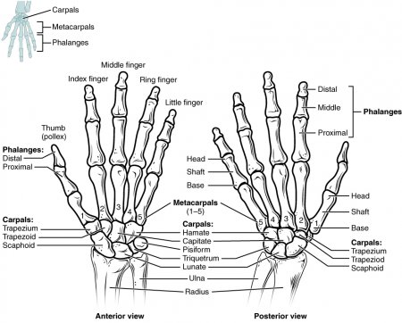
The bones of the hand and wrist provide the body with support and flexibility to manipulate objects in many different ways. Each hand contains 27 distinct bones that give the hand an incredible range and precision of motion. The forearm's ulna and radius support the many muscles that manipulate the bones of the hand and wrist. Rotation of the radius around the ulna results in the supination and pronation of the hand. These bones also form the flexible wrist joint with the proximal row of the carpals.
The ulna
- Category: Musculoskeletal system
- Views: 117515
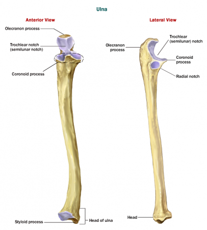
The ulna is the longer, larger and more medial of the lower arm bones. Many muscles in the arm and forearm attach to the ulna to perform movements of the arm, hand and wrist. Movement of the ulna is essential to such everyday functions as throwing a ball and driving a car.
The ulna extends through the forearm from the elbow to the wrist, narrowing significantly towards its distal end. At its proximal end it forms the elbow joint with the humerus of the upper arm and the radius of the forearm. The ulna extends past the humerus to form the tip of the elbow, known as the olecranon. The olecranon fits into a small recess in the humerus known as the olecranon fossa, preventing the elbow’s extension beyond around 180 degrees. Just distal to the olecranon is the concave trochlear notch that surrounds the trochlea of the humerus to form the hinge of the elbow joint. The distal lip of the trochlear notch protrudes anteriorly to form the coronoid process that helps to lock the ulna in place with the humerus at the elbow and fits into the coronoid fossa of the humerus. On the lateral edge of the coronoid process is the small radial notch that forms the proximal radioulnar joint with the radius and permits the radius to rotate around the ulna at the elbow. A long ridge on the anterior side of the coronoid process known as the ulnar tuberosity extends down the shaft of the ulna as a muscle attachment point.
The radius
- Category: Musculoskeletal system
- Views: 83679
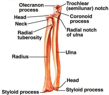
The radius is the more lateral and slightly shorter of the two forearm bones. It is found on the thumb side of the forearm and rotates to allow the hand to pivot at the wrist. Several muscles of the arm and forearm have origins and insertions on the radius to provide motion to the upper limb. These movements are essential to many everyday tasks such as writing, drawing, and throwing a ball.
The radius is located on the lateral side of the forearm between the elbow and the wrist joints. It forms the elbow joint on its proximal end with the humerus of the upper arm and the ulna of the forearm. Although the radius begins as the smaller of the two forearm bones at the elbow, it widens significantly as it extends along the forearm to become much wider than the ulna at the wrist. A short cylinder of smooth bone forms the head of the radius where it meets the capitulum of the humerus and the radial notch of the ulna at the elbow. The head of the radius allows the forearm to flex and pivot at the elbow joint. Just distal to the head, the radius narrows considerably to form the neck of the radius before expanding medially to form the radial tuberosity, a bony process that serves as the insertion of the biceps brachii.
The subdeltoid bursa
- Category: Musculoskeletal system
- Views: 86633
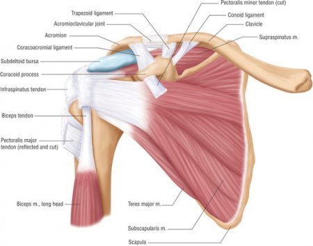
The subdeltoid bursa is a fluid-filled sac located under the deltoid muscle in the shoulder joint. It plays an important role in decreasing friction in the shoulder joint and protects the surrounding tissues of the joint.
The humerus
- Category: Musculoskeletal system
- Views: 95591
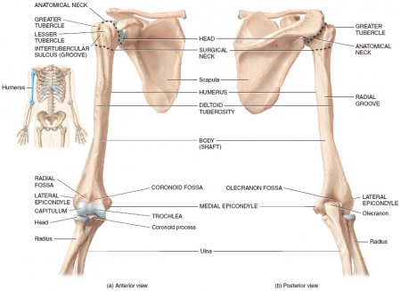
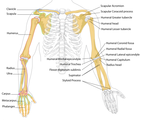
The humerus is the both the largest bone in the arm and the only bone in the upper arm. Many powerful muscles that manipulate the upper arm at the shoulder and the forearm at the elbow are anchored to the humerus. Movement of the humerus is essential to all of the varied activities of the arm, such as throwing, lifting, and writing.
At its proximal end, the humerus forms a smooth, spherical structure known as the head of the humerus. The head of the humerus forms the ball of the ball-and-socket shoulder joint, with the glenoid cavity of the scapula acting as the socket. The rounded shape of the head of the humerus allows the humerus to move in a complete circle (circumduction) and rotate around its axis at the shoulder joint. Just below the head, the humerus narrows into the anatomical neck of the humerus. Two small processes, the greater and lesser tubercles, extend from the humerus just below the anatomical neck as attachment points for the muscles of the rotator cuff. The humerus narrows below the tubercles again in a region known as the surgical neck before extending toward elbow joint as the body of the humerus. About a third of the way to the elbow, the humerus swells into a small process known as the deltoid tuberosity, which supports the insertion point of the deltoid muscle.
The scapula
- Category: Musculoskeletal system
- Views: 85798
The scapula is the technical name for the shoulder blade. It is a flat, triangular bone that lies over the back of the upper ribs. The rear surface can be felt under the skin. It serves as an attachment for some of the muscles and tendons of the arm, neck, chest and back and aids in the movements of the arm and shoulder. It is well padded with muscle so that great force is required to fracture it. The back surface of each scapula is divided into unequal portions by a spine. This spine leads to a head, which bears two processes-the acromion process that forms the tip of the shoulder and a coracoid process that curves forward and down below the clavicle (collarbone). The acromion process joins a clavicle and provides attachments for muscles of the arm and chest muscles. The acromion is a bony prominence at the top of the shoulder blade. On the head of the scapula, between the processes mentioned above, is a depression called the glenoid cavity. It joins with the head of the upper arm bone (humerus).
The clavicle (collarbone)
- Category: Musculoskeletal system
- Views: 82136

The clavicles, or collarbones, are a pair of long bones that connect the scapula to the sternum. The name clavicle comes from the Latin word for “little key” and describes the shape of the clavicle as an old-fashioned skeleton key. The clavicle is one of the most commonly broken bones in the human body. It also serves as an important and easily located bony landmark due to its superficial location and projection from the trunk.
The bones of the arm and hand
- Category: Musculoskeletal system
- Views: 8146
The bones of the arm and hand have the important jobs of supporting the upper limb and providing attachment points for the muscles that move the upper limb. These bones form joints that provide a wide range of motion and flexibility needed to manipulate objects deftly with the arm and hand. They also provide strength to resist the extreme forces and stresses acting upon the arms and hands during sports, exercise, and heavy labor.
Consisting of the clavicle (collar bone) and scapula (shoulder blade), the pectoral girdle forms the attachment point between the arm and the chest. The clavicle, which gets its name from the Latin word for key, is a long bone that connects the scapula to the sternum (breast bone) of the chest. It is located just under the skin in the thoracic region between the shoulder and the base of the neck. The clavicle is slightly curved like a letter S and is about five inches in length. Two joints are formed by the clavicle – the sternoclavicular joint with the sternum, and the acromioclavicular joint with the acromion of the scapula. The clavicle permits the shoulder joint to move in circles while remaining attached to the bones of the chest.
The coccyx
- Category: Musculoskeletal system
- Views: 81506
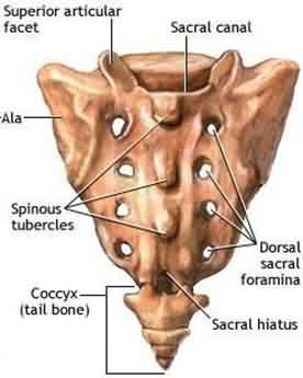
The coccyx, or tailbone, is both the smallest and the most inferior bone in the spinal column. It is a vestige of the caudal vertebrae found in the tails of most mammals. In the human body, the coccyx functions to anchor several muscles of the pelvic region and acts as one of the bones that bear the body’s weight while sitting.
The coccyx is a small, triangular bone measuring less than one inch (2.5 cm) in length and curved like a hawk’s beak. It is widest at its superior border, where it meets the sacrum, and is narrowest at its inferior tip. The coccyx is curved to form a concave surface anteriorly that is continuous with the curvature of the sacrum. In men the concavity is greater, while in females the concavity is reduced so that the coccyx does not point as anteriorly as it does in the male skeleton. A thin band of fibrocartilage binds the coccyx to the sacrum above it while permitting slight flexion and extension of the coccyx. Two bony spines known as cornua extend superiorly toward the sacrum on the posterior side and are connected to the sacral cornua by ligaments.
The ilium
- Category: Musculoskeletal system
- Views: 101538
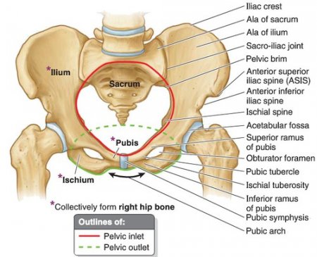
The ilium is the largest and most superior of the three bones that join to form the hipbone, or os coxa. It is a wide, flat bone that provides many attachment points for muscles of the trunk and hip. You can find the crest of your ilium by placing your hands on your hips. The superficial location of the ilium makes it a common site for extracting bone tissue for grafting and bone marrow for transplants.
