The stomach
- Category: Digestive system
- Views: 102336
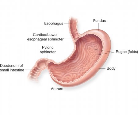
The stomach is the main food storage tank of the body. If it were not for the stomach’s storage capacity, we would have to eat constantly instead of just a few times each day. The stomach also secretes a mixture of acid, mucus, and digestive enzymes that helps to digest and sanitize our food while it is being stored.
Anatomy of the stomach
Gross Anatomy
The stomach is a rounded, hollow organ located just inferior to the diaphragm in the left part of the abdominal cavity. Located between the esophagus and the duodenum, the stomach is a roughly crescent-shaped enlargement of the gastrointestinal tract. The inner layer of the stomach is full of wrinkles known as rugae (or gastric folds). Rugae both allow the stomach to stretch in order to accommodate large meals and help to grip and move food during digestion.
The esophagus
- Category: Digestive system
- Views: 95538
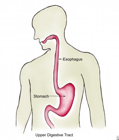
The esophagus is a long, thin, and muscular tube that connects the pharynx (throat) to the stomach. It forms an important piece of the gastrointestinal tract and functions as the conduit for food and liquids that have been swallowed into the pharynx to reach the stomach.
The esophagus is about 9-10 inches (25 centimeters) long and less than an inch (2 centimeters) in diameter when relaxed. It is located just posterior to the trachea in the neck and thoracic regions of the body and passes through the esophageal hiatus of the diaphragm on its way to the stomach.
At the superior end of the esophagus is the upper esophageal sphincter that keeps the esophagus closed where it meets the pharynx. The upper esophageal sphincter opens only during the process of swallowing to permit food to pass into the esophagus. At the inferior end of the esophagus, the lower esophageal sphincter opens for the purpose of permitting food to pass from the esophagus into the stomach. Stomach acid and chyme (partially digested food) is normally prevented from entering the esophagus, thanks to the lower esophageal sphincter. If this sphincter weakens, however, acidic chyme may return to the esophagus in a condition known as acid reflux. Acid reflux can cause damage to the esophageal lining and result in a burning sensation known as heartburn. If these symptoms occur with enough frequency, they are known as GERD (gastroesophageal reflux disease).
Physiology of the liver
- Category: Digestive system
- Views: 11112
Digestion
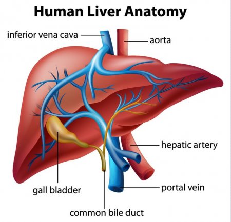
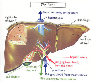
The liver plays an active role in the process of digestion through the production of bile. Bile is a mixture of water, bile salts, cholesterol, and the pigment bilirubin. Hepatocytes in the liver produce bile, which then passes through the bile ducts to be stored in the gallbladder. When food containing fats reaches the duodenum, the cells of the duodenum release the hormone cholecystokinin to stimulate the gallbladder to release bile. Bile travels through the bile ducts and is released into the duodenum where it emulsifies large masses of fat. The emulsification of fats by bile turns the large clumps of fat into smaller pieces that have more surface area and are therefore easier for the body to digest.
Bilirubin present in bile is a product of the liver’s digestion of worn out red blood cells. Kupffer cells in the liver catch and destroy old, worn out red blood cells and pass their components on to hepatocytes. Hepatocytes metabolize hemoglobin, the red oxygen-carrying pigment of red blood cells, into the components heme and globin. Globin protein is further broken down and used as an energy source for the body. The iron-containing heme group cannot be recycled by the body and is converted into the pigment bilirubin and added to bile to be excreted from the body. Bilirubin gives bile its distinctive greenish color. Intestinal bacteria further convert bilirubin into the brown pigment stercobilin, which gives feces their brown color.
The liver
- Category: Digestive system
- Views: 101599
Weighing in at around 3 pounds, the liver is the body’s second largest organ; only the skin is larger and heavier. The liver performs many essential functions related to digestion, metabolism, immunity, and the storage of nutrients within the body. These functions make the liver a vital organ without which the tissues of the body would quickly die from lack of energy and nutrients. Fortunately, the liver has an incredible capacity for regeneration of dead or damaged tissues; it is capable of growing as quickly as a cancerous tumor to restore its normal size and function.
The epiglottis
- Category: Digestive system
- Views: 92423
The epiglottis is a flexible flap at the superior end of the larynx in the throat. It acts as a switch between the larynx and the esophagus to permit air to enter the airway to the lungs and food to pass into the gastrointestinal tract. The epiglottis also protects the body from choking on food that would normally obstruct the airway.
The epiglottis is a thin, leaf-shaped structure at the superior border of the larynx, or voice box. In its relaxed position, the epiglottis projects into the pharynx, or throat, and rests just posterior to the tongue. Viewed from the posterior direction, it is shaped like a teardrop with a wide, rounded region at the superior end and a narrow tapered point at its inferior end. The epiglottis is also concave with the lateral edges pointing posteriorly. Two tiny ligaments - the thyroepiglottic and hyoepiglottic ligaments - hold the epiglottis in its resting position in the throat. The thin thyroepiglottic ligament connects the inferior point of the epiglottis to the thyroid cartilage of the larynx, while the hyoepiglottic ligament connects the anterior surface of the superior region to the hyoid bone.
The teeth
- Category: Digestive system
- Views: 8141
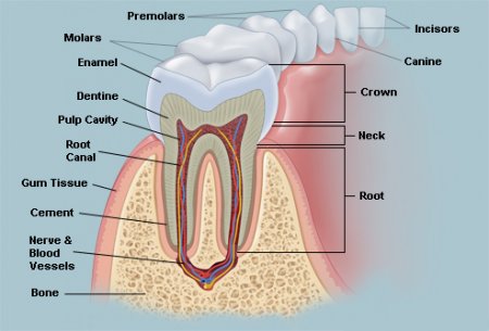
By the shape the teeth are divided into:
- incisors - have one root, wedge-shaped crowns, particularly upper incisors crown shape has the spatula, lower - bits;
- canines - have one root, crown conical shape;
- premolars: upper premolars have bifurcated root, on the horizontal cut their crown has oval shape; hills are almost identical; lower premolars have one root, on the horizontal cut their crown has round shape, vestibular hill is large, oral - smaller;
- molars: upper molars have three roots (two vestibular and one oral), diamond-shaped crown; lower molars have two roots, square shape of crown.
Adult human has 28-32 permanent teeth. Child has 20 milk teeth.
The teeth are located in the mouth.
The salivary glands
- Category: Digestive system
- Views: 8141
Salivary glands produce saliva.
Salivary glands are divided into small and large.
Small salivary glands located in the mucous membrane of the mouth:
- labial;
- buccal;
- palatine;
- lingual;
- molar.
Large salivary glands:
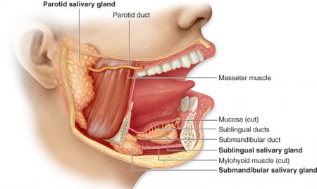
1) parotid gland - paired, is located near the ear, in the mandibular fold. Duct of parotid gland located on chewing muscle, passes through the buccal muscle and opens in the cheek mucosa opposite of 2nd upper molar. Parotid salivary gland - parenchymal organ, anatomical unit of which is the a particle;
2) submandibular gland - paired, located in submandibular triangle. Duct of submandibular gland opens in the proper oral cavity in the sublingual meet. Submandibular gland - parenchymal organ, anatomical unit of which is the a particle;
3) sublingual gland - paired, located in the sublingual crease. Large duct of sublingual gland opens in the sublingual meet, small duct opens directly in sublingual crease. Sublingual gland - parenchymal organ, anatomical unit of which is a particle.
The tongue
- Category: Digestive system
- Views: 23191
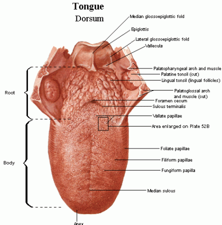
The tongue has a conical shape. The tongue - unpaired body. The tongue is located in the oral cavity.
The outer structure of the tongue
Tongue is divided:
- root of the tongue (radix linguae);
- body of the tongue (corpus linguae);
- apex (tip) of the tongue (apex linguae);
- the back of the tongue (dorsum linguae);
- the lower surface of the tongue (facies inferior linguae).
On the back of the tongue is a blind hole (foramen cecum linguae). From the blind hole aside and ahead goes steam frontier furrow. This furrow divides the tongue into pre-furrow part and after-furrow part.
The internal structure of the tongue
The tongue consists of muscles covered with mucous membrane. The muscles of the tongue are divided into skeletal and own. Skeletal muscle:
- Awl-lingual muscle - starts from subulate process, shifts the tongue up and back;
- Sublingual-lingual muscle - starts from the hyoid bone, the tongue shifts back and down; aside;
- Chin-lingual muscle - starts from chin spine, shifts the tongue ahead.
Development of the oral cavity
- Category: Digestive system
- Views: 7121
In the main part of anterior part of primary intestine develops gill apparatus, which consists of 5 pairs of gill pockets and gill arches. Gill pockets - a protrusion of lateral walls endoderm of the primary intestine. Gill arches - a section of mesenchyme placed between adjacent gill pockets. Primary oral cavity (mouth bay) has the form of cracks, bounded with 5 processes, derivatives of the first pair of gill arches. The upper edge of the mouth bay bounded by unpaired frontal process and by two maxillary processes, the lower edge - by two mandibular processes. Mandibular grow, processes and form the lower jaw, soft parts of the face, chin, lower lip. If mandibular processes do not grow, arises an anomaly - midpoint separation of the lower jaw, lips. Maxillary processes form the upper jaw, palate, lateral parts of the upper lip, cheeks.
Oral cavity
- Category: Digestive system
- Views: 103863
Mouth has 2 parts:
1) oral vestibule
2) proper oral cavity.
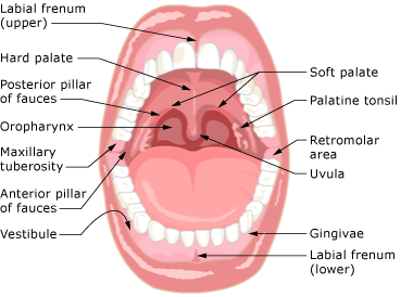
1. Vestibule of oral cavity is the space bounded by the upper and lower lips, cheeks (front) and also by teeth and gums (rear).The upper lip is bounded by basis of nose and pair nasolabial sulcus; lower lip is bounded by chin-lip sulcus. Lips are formed by circular muscle of the mouth, which is covered with skin on the outside and by mucous membrane on inside. There are skin part of the lip, intermediate - red border, mucous. In the transition from the upper lip to the alveolar bone of the upper jaw mucous membrane forms a fold - the bridle of upper lip; in the transition from the alveolar part of lower lip to the lower jaw mucous membrane forms a fold - bridle of lower lip. Between the lips is oral cleft. Cheek is bounded in above by zygomatic arch, in bottom – by edge of the lower jaw, in front – by nasolabial sulcus, in behind - by chewing muscles. The basis of cheek is buccal muscle lined with mucous membrane inside, and the outside is covered with skin. In cheek is pronounced subcutaneous fat, which forms the fat body of cheeks.
2. Proper oral cavity is bounded by the upper and lower walls. The upper wall is a palate. There are:
- hard palate, formed by palate bone and mucous membrane that covers the bone palate;
- soft palate, formed by muscles and mucous membrane that covers the muscles.
