The small intestine
- Category: Digestive system
- Views: 83917
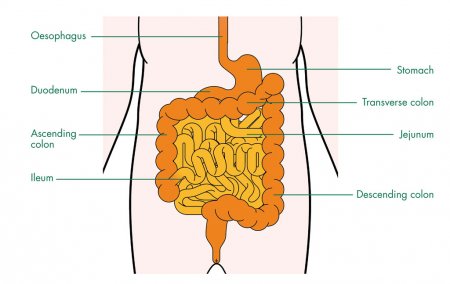
The small intestine is a long, highly convoluted tube in the digestive system that absorbs about 90% of the nutrients from the food we eat. It is given the name “small intestine” because it is only 1 inch in diameter, making it less than half the diameter of the large intestine. The small intestine is, however, about twice the length of the large intestine and usually measures about 10 feet in length.
The small intestine winds throughout the abdominal cavity inferior to the stomach. Its many folds help it to pack all 10 feet of its length into such a small body cavity. A thin membrane known as the mesentery extends from the posterior body wall of the abdominal cavity to surround the small intestine and anchor it in place. Blood vessels, nerves, and lymphatic vessels pass through the mesentery to support the tissues of the small intestine and transport nutrients from food in the intestines to the rest of the body.
The spleen
- Category: Digestive system, Immune and lymphatic systems
- Views: 156767
The spleen is a brown, flat, oval-shaped lymphatic organ that filters and stores blood to protect the body from infections and blood loss.
Protected by our ribs, the spleen is located between the stomach and the diaphragm in the left hypochondriac region of the abdominal body cavity. The splenic artery branches off from the aorta and the celiac trunk to deliver oxygenated blood to the spleen, while the splenic vein carries deoxygenated blood away from the spleen to the hepatic portal vein. A tough connective tissue capsule surrounds the soft inner tissue of the spleen.
Physiology of the pancreas
- Category: Digestive system, Endocrine system
- Views: 9319
Digestion
The exocrine portion of the pancreas plays a major role in the digestion of food. The stomach slowly releases partially digested food into the duodenum as a thick, acidic liquid called chyme. The acini of the pancreas secrete pancreatic juice to complete the digestion of chyme in the duodenum. Pancreatic juice is a mixture of water, salts, bicarbonate, and many different digestive enzymes. The bicarbonate ions present in pancreatic juice neutralize the acid in chyme to protect the intestinal wall and to create the proper environment for the functioning of pancreatic enzymes. The pancreatic enzymes each specialize in digesting specific compounds found in chyme.
- Pancreatic amylase breaks large polysaccharides like starches and glycogen into smaller sugars such as maltose, maltotriose, and glucose. Maltase secreted by the small intestine then breaks maltose into the monosaccharide glucose, which the intestines can directly absorb.
- Trypsin, chymotrypsin, and carboxypeptidase are protein-digesting enzymes that break proteins down into their amino acid subunits. These amino acids can then be absorbed by the intestines.
- Pancreatic lipase is a lipid-digesting enzyme that breaks large triglyceride molecules into fatty acids and monoglycerides. Bile released by the gallbladder emulsifies fats to increase the surface area of triglycerides that pancreatic lipase can react with. The fatty acids and monoglycerides produced by pancreatic lipase can be absorbed by the intestines.
- Ribonuclease and deoxyribonuclease are nucleases, or enzymes that digest nucleic acids. Ribonuclease breaks down molecules of RNA into the sugar ribose and the nitrogenous bases adenine, cytosine, guanine and uracil. Deoxyribonuclease digests DNA molecules into the sugar deoxyribose and the nitrogenous bases adenine, cytosine, guanine, and thymine.
The pancreas
- Category: Digestive system, Endocrine system
- Views: 162629
The pancreas is a glandular organ in the upper abdomen, but really it serves as two glands in one: a digestive exocrine gland and a hormone-producing endocrine gland. Functioning as an exocrine gland, the pancreas excretes enzymes to break down the proteins, lipids, carbohydrates, and nucleic acids in food. Functioning as an endocrine gland, the pancreas secretes the hormones insulin and glucagon to control blood sugar levels throughout the day. Both of these diverse functions are vital to the body’s survival.
Physiology of the gallbladder
- Category: Digestive system
- Views: 12229
Storage
The gallbladder acts as a storage vessel for bile produced by the liver. Bile is produced by hepatocytes cells in the liver and passes through the bile ducts to the cystic duct. From the cystic duct, bile is pushed into the gallbladder by peristalsis (muscle contractions that occur in orderly waves). Bile is then slowly concentrated by absorption of water through the walls of the gallbladder. The gallbladder stores this concentrated bile until it is needed to digest the next meal.
Stimulation
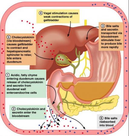
Foods rich in proteins or fats are more difficult for the body to digest when compared to carbohydrate-rich foods (see Macronutrients). The walls of the duodenum contain sensory receptors that monitor the chemical makeup of chyme (partially digested food) that passes through the pyloric sphincter into the duodenum. When these cells detect proteins or fats, they respond by producing the hormone cholecystokinin (CCK). CCK enters the bloodstream and travels to the gallbladder where it stimulates the smooth muscle tissue in the walls of the gallbladder.
The gallbladder
- Category: Digestive system
- Views: 89235
The gallbladder is a small storage organ located inferior and posterior to the liver. Though small in size, the gallbladder plays an important role in our digestion of food. The gallbladder holds bile produced in the liver until it is needed for digesting fatty foods in the duodenum of the small intestine. Bile in the gallbladder may crystallize and form gallstones, which can become painful and potentially life threatening.
Physiology of the stomach
- Category: Digestive system
- Views: 15683
Storage
In the mouth, we chew and moisten solid food until it becomes a small mass known as a bolus. When we swallow each bolus, it then passes through the esophagus to the stomach where it is stored along with other boluses and liquids from the same meal.
The size of the stomach varies from person to person, but on average it can comfortably contain 1-2 liters of food and liquid during a meal. When stretched to its maximum capacity by a large meal or overeating, the stomach may hold up to 3-4 liters. Distention of the stomach to its maximum size makes digestion difficult, as the stomach cannot easily contract to mix food properly and leads to feelings of discomfort.
The stomach
- Category: Digestive system
- Views: 102328
The stomach is the main food storage tank of the body. If it were not for the stomach’s storage capacity, we would have to eat constantly instead of just a few times each day. The stomach also secretes a mixture of acid, mucus, and digestive enzymes that helps to digest and sanitize our food while it is being stored.
Anatomy of the stomach
Gross Anatomy
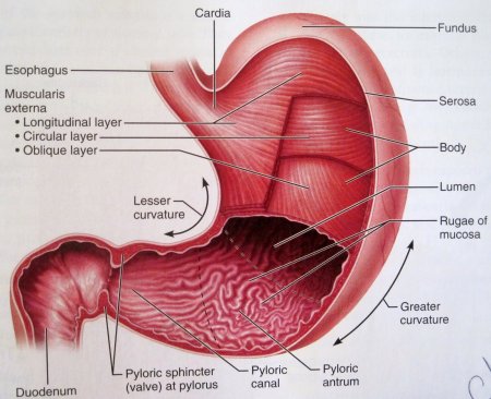
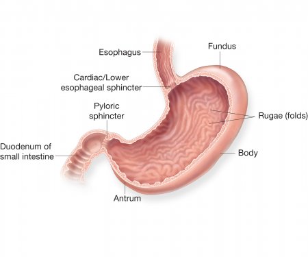
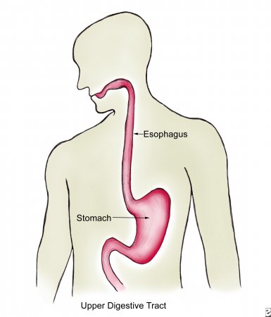
The stomach is a rounded, hollow organ located just inferior to the diaphragm in the left part of the abdominal cavity. Located between the esophagus and the duodenum, the stomach is a roughly crescent-shaped enlargement of the gastrointestinal tract. The inner layer of the stomach is full of wrinkles known as rugae (or gastric folds). Rugae both allow the stomach to stretch in order to accommodate large meals and help to grip and move food during digestion.
The esophagus
- Category: Digestive system
- Views: 95523
The esophagus is a long, thin, and muscular tube that connects the pharynx (throat) to the stomach. It forms an important piece of the gastrointestinal tract and functions as the conduit for food and liquids that have been swallowed into the pharynx to reach the stomach.
The esophagus is about 9-10 inches (25 centimeters) long and less than an inch (2 centimeters) in diameter when relaxed. It is located just posterior to the trachea in the neck and thoracic regions of the body and passes through the esophageal hiatus of the diaphragm on its way to the stomach.
At the superior end of the esophagus is the upper esophageal sphincter that keeps the esophagus closed where it meets the pharynx. The upper esophageal sphincter opens only during the process of swallowing to permit food to pass into the esophagus. At the inferior end of the esophagus, the lower esophageal sphincter opens for the purpose of permitting food to pass from the esophagus into the stomach. Stomach acid and chyme (partially digested food) is normally prevented from entering the esophagus, thanks to the lower esophageal sphincter. If this sphincter weakens, however, acidic chyme may return to the esophagus in a condition known as acid reflux. Acid reflux can cause damage to the esophageal lining and result in a burning sensation known as heartburn. If these symptoms occur with enough frequency, they are known as GERD (gastroesophageal reflux disease).
Physiology of the liver
- Category: Digestive system
- Views: 11110
Digestion
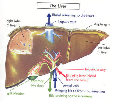
The liver plays an active role in the process of digestion through the production of bile. Bile is a mixture of water, bile salts, cholesterol, and the pigment bilirubin. Hepatocytes in the liver produce bile, which then passes through the bile ducts to be stored in the gallbladder. When food containing fats reaches the duodenum, the cells of the duodenum release the hormone cholecystokinin to stimulate the gallbladder to release bile. Bile travels through the bile ducts and is released into the duodenum where it emulsifies large masses of fat. The emulsification of fats by bile turns the large clumps of fat into smaller pieces that have more surface area and are therefore easier for the body to digest.
Bilirubin present in bile is a product of the liver’s digestion of worn out red blood cells. Kupffer cells in the liver catch and destroy old, worn out red blood cells and pass their components on to hepatocytes. Hepatocytes metabolize hemoglobin, the red oxygen-carrying pigment of red blood cells, into the components heme and globin. Globin protein is further broken down and used as an energy source for the body. The iron-containing heme group cannot be recycled by the body and is converted into the pigment bilirubin and added to bile to be excreted from the body. Bilirubin gives bile its distinctive greenish color. Intestinal bacteria further convert bilirubin into the brown pigment stercobilin, which gives feces their brown color.
