The digestive system provides receiving, mechanical and chemical processing of food, products absorption of splitting and removal of undigested residues.
The organs of the digestive system include digestive channel through which food passes (mouth, esophagus, stomach, intestines) and digestive glands (salivary, pancreas, liver, etc.). The wall of the digestive tract consists of three layers: the outer, middle and inner. The outer layer is formed by connective tissue that separates the digestive tube from surrounding tissues and organs. The middle layer - muscular. In the upper sections muscle layer is formed by striated and in the middle of the esophagus - by smooth muscle tissue. The inner layer - mucosa.
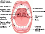
Digestive tract begins with oral cavity (cavitas oris). It is formed by lips, cheeks, palate, tongue and oral cavity of the bottom muscles. The walls of oral cavity are lined with mucous membrane that contains numerous small glands that secrete saliva.
Two rows of teeth (dentes) divide oral cavity to front mouth cavity and mouth. Teeth - bone formation, used for grinding of food. Human teeth with other bodies involved in sound formation.
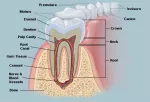
Rudiments of teeth laid during embryonic development. Approximately from 5-6 month after the birth develops the first generation of teeth - milk teeth that from 6 years begin to be replaced with permanent teeth. Adult human has total 32 teeth: 8 incisors, 4 canines, 8 small and 12 large roots. They differ in structure and function. The tooth consists of the top, or crown, neck and root. The bulk of the tooth include dentin, in the crown area tooth is covered with enamel, in the neck (in mammals) - with cement. Inside the tooth is cavity - root canal filled with tooth flesh or pulp.
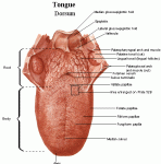
The tongue of man (and other mammals) is formed with striated muscle tissue, covered with mucous membrane, in which are located taste buds. The tongue performs many different functions: participation in the chewing, swallowing, speech articulation; tongue is the organ of taste. The tongue of human children (and mammals cubs) provides an extremely important role in the sucking of mother\'s milk. The tongue consists of a root, body and top. At the top of the tongue are located receptors that perceive sweet taste, the sides of the tongue - salty and sour, the roots - bitter. With receptors person feels also mechanical properties and temperature of the food. In oral cavity open ducts of three pairs of large salivary glands: parotid, submandibular and sublingual.
Behind the oral cavity is located pharynx. It is wide tube length of about 6 inches, flattened in the anteroposterior direction that narrows at the transition into the esophagus. Pharyngeal wall consists of an inner layer - the mucosa, which is covered with ciliated epithelium in the field of nasal and multilayered - in the mouth and throat parts, and a layer of striated muscles. At the level of the 6th cervical vertebra the throat turns into the esophagus.
The esophagus is cylindrical muscular tube length of 9-10 inches. The upper third of the esophagus is composed of striated muscle, and the rest of the length - two layers of smooth muscle: the outer - longitudinal and internal - ring. In front the esophagus adjacent to the trachea. The muscles of the esophagus, by contraction, push food into the stomach.
Single-chamber stomach (ventriculus, gaster) - expanded part of the digestive canal in volume of 1.5-2 liters. Stomach form and capacity depends on the characteristics of the constitution and can vary in the same person. The stomach can take the form of long curved horns or bag. In stomach are distinguished small (upper) and large (lower) curvature. There are also the following parts of the stomach: the top - bottom, middle - body and lower - pylorus. In the stomach wall are three main groups of glands: the main that secrete pepsin and chymosin; overlaying that secrete hydrochloric acid; additional that secrete mucus. The mucous membrane forms folds. The muscles of the stomach wall consists of three layers: longitudinal, ring and oblique. In place of transition of the stomach into the duodenum annular layer thickens and forms a sphincter.
After the stomach is located small intestine (intestinum tenue) length of 5-7 meters. It consists of the duodenum, empty and tangle of intestines. The wall of the small intestine include the following layers: mucous, muscular and serous. The mucous membrane has a huge number (up to 30 million) of microscopic outgrowths - villous height of 0,3- 1,2 millimeters, which increase sucking surface of the small intestine in 1000 times. Between the main cells of this membrane, acting as suction function are located goblet cells which secrete mucus. Muscle membrane is composed of the small intestine of smooth muscles, they create internal (circular) and external (longitudinal) layers. Thickness of this layers is much less than the thickness of the stomach wall. Serous membrane covering also the whole small intestine, and create ripples of the small intestine in which located vessels and nerves.
The initial section of the small intestine - duodenum has a length of 10 inches, with diameter - 2 inches, Duodenum has a form of the horseshoe bend, It has open ducts of the liver and pancreas. The diameter of the jejunum is 1.5-2.5 inches, iliac (ileum) - 1,5-2,0 inches. Glands of the small intestine wall secrete intestinal juice that is the turbid viscous liquid. During the day is secrete around of 2 liters of intestinal juice. Reaction of the small intestine environment is alkaline: in it neutralized acidic environment of stomach contents that comes here. Intestinal juice contains more than 20 enzymes that cleave proteins, fats, carbohydrates and nucleic acids, and enterokinase enzyme which converts inactive trypsinogen to active trypsin.
Behind the stomach in the duodenum bend is located pancreas. The length of 5-6 inches. Pancreas consists of a head, body, tail and has a lobed structure. Along the whole gland passes duct through which pancreatic juice is secreted into the duodenum. Pancreatic juice carries alkaline reaction. It contains enzymes that break down proteins (proteases), fats (lipase), carbohydrates (amylase and maltose) and nucleic acids (nucleases). Pancreas - gland of mixed secretions because its special cells produce hormones that regulate carbohydrate metabolism.
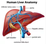
Liver (hepar) - the largest digestive gland of human organism, its weight of 1,5-2 kg. It is located mainly in the right upper quadrant, under the diaphragm. The upper surface is convex, lower is little concave. In the liver are four unequal parts. The largest part is the right part, lies in right upper quadrant. At the bottom of the liver, in the center, are located liver gates through which pass the blood vessels, nerves and bile ducts. In the deepening on the bottom surface is located the gallbladder (vesica fellea) with volume of 40-70 ml. The liver is covered with peritoneum. With connection it is kept in a certain position. The basic structural and functional unit of the liver is the liver particles, which form the parts. The liver secretes per day from 500 to 1200 ml of bile. Bile is secretes continuously and flow to the intestine with food. Bile is a yellow liquid. It consists of water, acids and bile pigments, cholesterol, mineral salts. Through the common bile duct bile secretes to the duodenum.
The small intestine goes into large intestine (intestinum crassum) length of 1,5-2 m. It is a larger diameter (4-8 cm.), and therefore so named. The large intestine include cecum (caecum) with appendix, colon, sigmoid and rectum. Rectum ends with the anus (anus). Large intestine mucous membrane forms folds. It is lined with a single layer of cylindrical epithelium, villi are absent. The muscular layer of the large intestine is much larger than the small intestine. In it secrete intestinal juice that has an alkaline reaction and poor in enzymes. In this intestine section is a huge number of microorganisms, among which dominates escherichia coli.


