The muscles of the abdomen, lower back, and pelvis are separated from those of the chest by the muscular wall of the diaphragm, the critical breathing muscle. Lying exposed between the protective bones of the superiorly located ribs and the inferiorly located pelvic girdle, the muscles of this region play a critical role in protecting the delicate vital organs within the abdominal cavity. In addition to providing protection, these core muscles also function in movement of the trunk, posture, and stability of the entire body.
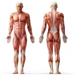
Extending across the anterior surface of the body from the superior border of the pelvis to the inferior border of the ribcage are the muscles of the abdominal wall, including the transverse and rectus abdominis and the internal and external obliques. Working as a team, these muscles contract to flex, laterally bend, and rotate the torso. The abdominal muscles also play a major role in the posture and stability to the body and compress the organs of the abdominal cavity during various activities such as breathing and defecation.
The muscles of the lower back, including the erector spinae and quadratus lumborum muscles, contract to extend and laterally bend the vertebral column. These muscles provide posture and stability to the body by holding the vertebral column erect and adjusting the position of the body to maintain balance.
Attached to the pelvis are muscles of the buttocks, the lower back, and the thighs. These muscles, including the gluteus maximus and the hamstrings, extend the thigh at the hip in support of the body\'s weight and propulsion. Other pelvic muscles, such as the psoas major and iliacus, serve as flexors of the trunk and thigh at the hip joint and laterally rotate the hip as well.
The external abdominal obliques muscle
The external abdominal obliques are a pair of broad, thin, superficial muscles that lie on the lateral sides of the abdominal region of the body. Contraction of these muscles may result in several different actions, but they are best known for their lateral flexion and rotation of the trunk known as a side bend. The external obliques get their name from their position in the abdomen external to the internal abdominal obliques and from the direction of their fibers, which run obliquely (diagonally) across the sides of the abdomen.
The external abdominal obliques have their origins along the lateral ribs 5 through 12 and insert into the linea alba of the abdomen, the pubis, and the iliac crest of the hip bones. Their shape is roughly rectangular with the long axis running anterior to posterior along the linea alba. Muscle fibers in the external obliques run medially and inferiorly from the origins to the insertions across the lateral sides of the abdomen and end just lateral to the rectus abdominis muscles.
The location and structure of the external abdominal obliques gives them many different possible actions. Contraction of both external obliques together results in the compression of the abdomen (as in sucking in the gut) or the flexion of the trunk (as in performing a crunch or sit-up). Contraction of one of the abdominal obliques results in the lateral flexion and rotation of the trunk on the opposite side; in other words, the left external oblique rotates and flexes the trunk to the right.
The internal abdominal oblique muscle
The internal abdominal oblique muscle lies on the sides and front of the abdomen and is the intermediate of the three flat muscles in this area, below the external oblique and above the transverse abdominal muscle. It is broad, thin and irregularly four-sided and occupies the lateral walls of the abdomen, stretching across to the front. Both sides, acting together, flex the vertebral column by drawing the pubis toward the xiphoid process (the smallest of the three parts of the breastbone). One side also bends the vertebral column sideways and rotates it, bringing the shoulder of that side forward. The external abdominal oblique muscle is also irregularly four-sided in form and lies superficial to the internal oblique muscle. Both sides, acting together, flex the vertebral column, drawing cartilage down toward the pubis. One side acts alone bending the vertebral column sideways, rotating it to bring the shoulder of the opposite side forward. Both of the abdominal oblique muscles work to compress abdominal contents, assist in the digestive process and in forced expiration.
The rectus abdominis muscles
The rectus abdominis muscles, commonly referred to as the “abs,” are a pair of long, flat muscles that extend vertically along the entire length of the abdomen adjacent to the umbilicus. Each muscle consists of a string of four fleshy muscular bodies connected by narrow bands of tendon, which give it a lumpy appearance when well defined and tensed. This lumpy appearance results in the rectus abdominis muscles being referred to as the “six-pack.”
The name rectus abdominis comes from the Latin words for “straight” and “abdominal,” indicating that its fibers run in a straight vertical line through the abdominal region of the body. The rectus abdominis has its origins along the superior edge of the pubis bone and the pubic symphysis in the pelvis. Its insertions are at the inferior edges of the costal cartilages of the fifth through seventh ribs and at the xiphoid process of the sternum. A covering of connective tissue known as the rectus sheath surrounds the rectus abdominis muscles and provides attachment points for the internal and external oblique muscles that flank them on both sides. Between the rectus abdominis muscles is a thick mass of white fibrous connective tissue called the linea alba that unites the abdominal muscles of the left and right sides.
The rectus abdominis muscle performs the important task of flexing the torso and spine in the abdominal region. It does this by pulling the ribcage closer to the pelvis. The rectus abdominis can also tense to contract the abdomen without moving the torso, as in sucking in one’s gut. Contraction of the abdomen results in increased pressure within the abdominopelvic cavity and is useful to push substances out of the body during exhalation, defecation, and urination.
The thoracolumbar fascia
The thoracolumbar fascia is a large portion of deep fascia in three parts found between the muscles of the back. As with any individual skeletal muscle, the muscles of the back are separated from adjacent muscles and held in place by layers of fibrous connective tissues called fascia. This connective tissue surrounds each muscle and may project beyond the end of its muscle fibers to form a cordlike tendon. Fibers in a tendon intertwine to attach the fibers of the muscle to the bone. The fascia associated with the individual organs of the muscular system is part of a complete network of fascia that extends throughout the body. The part of this network that surrounds and penetrates the muscles is called deep fascia. It is continuous with the subcutaneous fascia that forms the fascia covering organs in various body cavities as well as the cavities themselves.


