The facial nerve is one of the twelve pairs of cranial nerves in the peripheral nervous system. It is the seventh cranial nerve, and so is often referred to as cranial nerve VII or simply CN VII. Nerve signals from the cranial nerve play important roles in sensing taste as well as controlling the muscles of the face, salivary glands, and lacrimal glands.
Anatomy of the facial nerve
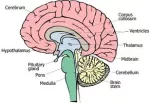
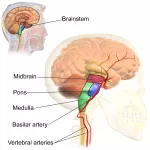
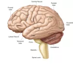
The facial nerve is the seventh cranial nerve to exit the brain when counting from anterior to posterior. It arises from the pons region of the brainstem, posterior to the abducens nerve (CN VI) and anterior to the vestibulocochlear nerve (CN VIII). The facial nerve travels from the pons through the facial canal in the temporal bone to exit the skull at the stylomastoid foramen.
As the facial nerve passes through the temporal bone, several smaller nerves branch off from the main nerve, including the greater (superficial) petrosal nerve and the chorda tympani.
- Nerve fibers from the greater (superficial) petrosal nerve stimulate the lacrimal glands to produce tears and moisten the eyes.
- The chorda tympani stimulates the submandibular and sublingual salivary glands to produce saliva. It also carries taste information from the anterior two-thirds of the tongue to the brain.
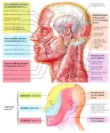
After passing through the stylomastoid foramen, the facial nerve emerges just inferior to the ear and splits into several superficial branches.
- The posterior auricular nerve splits from the facial nerve just beyond the stylomastoid foramen and innervates the muscles posterior to the ear, including the auricularis posterior and the occipitalis.
- Two small nerves next branch off to innervate the digastric and stylohyoid muscles.
- Finally, the temporofacial and cervicofacial branches separate to innervate the muscles of the upper and lower face, respectively. The temporofacial nerve divides into the temporal, zygomatic, and infraorbital branches to reach the frontalis and orbicularis oculi muscles, among others. Fibers from the cervicofacial branch split into the buccal, mandibular, and cervical nerves to innervate the nasalis, zygomaticus major, buccinator, orbicularis oris, platysma, and other muscles surrounding the nose and mouth.
Physiology of the facial nerve
The facial nerve is considered a mixed nerve because it contains both afferent (sensory) and efferent (motor) neurons. Afferent neurons of the facial nerve carry taste sensations from the taste buds of the anterior tongue to the primary gustatory center of the cerebrum. The efferent division of the facial nerve contains both somatic (voluntary) motor neurons and autonomic (involuntary) motor neurons. Somatic motor neurons carry nerve signals to the skeletal muscles of the face to control facial expressions, while autonomic motor neurons carry signals to the lacrimal and salivary glands.
Histology of the facial nerve
Like all nerves, the facial nerve is made of thousands of individual neurons bundled together with connective tissue and blood vessels. The outside of the nerve is covered in a layer of connective tissue known as the epineurium that protects the soft neurons within and holds the nerve together as a cohesive mass. Within the epineurium, small arterioles and venules provide blood flow to many small bundles of neurons known as fascicles. Each fascicle is wrapped in yet another layer of connective tissue known as the perineurium. Within each fascicle are several individual neurons wrapped in individual layers of connective tissue known as endoneurium.


