The nerves of the leg and foot
- Category: Nervous system
- Views: 113010
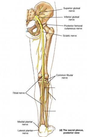
The nerves of the leg and foot serve to propel the body through the actions of the legs, feet, and toes while maintaining balance, both while the body is moving and when it is at rest. Sensory nerves are of course present throughout the lower extremities; however, with the exception of the bottom of the foot, they play a lesser role here than in the upper extremities. Primarily, this section of the peripheral nervous system sends and receives signals regarding locomotion and balance of the body.
The nerves of the arm and hand
- Category: Nervous system
- Views: 92499
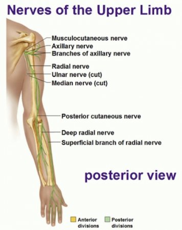
The nerves of the arm and hand perform a substantial two-fold role: commanding the intricate movements of the arms all the way down to the dexterous fingers, while also receiving the vast sensory information supplied by the sensory nerves of the hands and fingers. The movements of the arms must be fast, precise, and strong to complete the diverse activities the body engages in throughout the day. Even the tiny hand muscles, which perform very delicate and precise movements, are driven by about 200,000 neurons. Rapid conduction of sensory nerve signals from the hands provides critical information to the brain and feedback during precise activities.
The nervous system of the abdomen, lower back, and pelvis
- Category: Nervous system
- Views: 93886
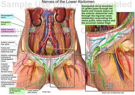
The nervous system of the abdomen, lower back, and pelvis contains many important nerve conduits that service this region of the body as well as the lower limbs. This section of the nervous system features the most inferior portion of the spinal cord along with many major nerves, plexuses, and ganglia that serve the vital organs of the abdominopelvic cavity.
The spinal cord
- Category: Nervous system
- Views: 84978
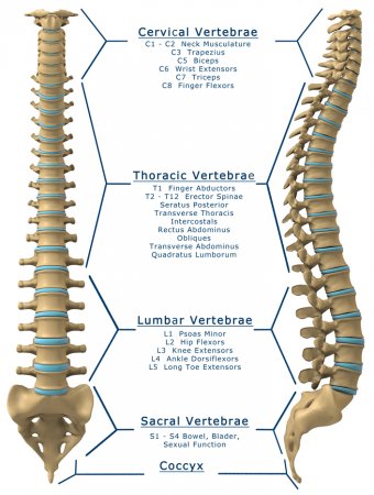
The spinal cord is a major component of the central nervous system (CNS) that forms the vital link between the brain and most of the body. Its 31 spinal segments connect the CNS to the organs and tissues of the neck, torso, and limbs. The spinal cord also performs important processing functions to maintain balance and respond quickly to stimuli.
Nerves of the chest and upper back
- Category: Nervous system
- Views: 84002
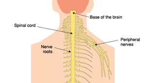
The nervous system of the thorax is a vital part of the nervous system as a whole, as it includes the spinal cord, peripheral nerves, and autonomic ganglia that communicate with and control many vital organs. Sensory information from the body and critical signals traveling to and from the limbs, trunk and vital organs all pass through this region on their way to and from the brain.
The taste buds
- Category: Nervous system
- Views: 87072
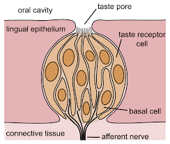
The taste buds are sensory taste receptors found on the tongue, throat, and palate that help form the perception of taste. Taste buds detect chemicals dissolved in saliva from food in the mouth and throat. Then, these taste buds send their sensory information through neurons to the gustatory center of the brain. The average person has around 10,000 taste buds in their mouth and throat, although the number of taste buds peaks in early childhood and declines throughout our lives.
The facial nerve
- Category: Nervous system
- Views: 94087
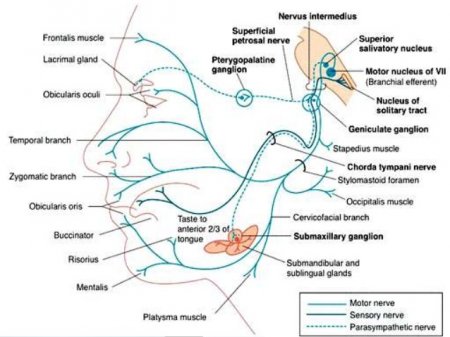
The facial nerve is one of the twelve pairs of cranial nerves in the peripheral nervous system. It is the seventh cranial nerve, and so is often referred to as cranial nerve VII or simply CN VII. Nerve signals from the cranial nerve play important roles in sensing taste as well as controlling the muscles of the face, salivary glands, and lacrimal glands.
The cranial nerve X - the vagus nerve
- Category: Nervous system
- Views: 84309
The tenth cranial nerve, and one of the most important, is the vagus nerve. All twelve of the cranial nerves, the vagus nerve included, emerge from or enter the skull (the cranium), as opposed to the spinal nerves which emerge from the vertebral column. The vagus nerve originates in the medulla oblongata, a part of the brain stem.
The eye's rods and cones
- Category: Nervous system
- Views: 83234
Rods and cones generate nerve impulses in the retinas of the eyes that travel along the optic nerves to the optic chiasma, where they partially cross over. The sensory organs for vision - the eyes - are at the front of the head, but areas of the brain at the back and sides provide the actual visual sense. Mixed impulses from both eyes pass through the optic tracts to the striate cortex at the back of the brain and end in the temporal lobe area so that right and left halves of the visual field merge. When light rays reach the retina (the film of the eye's camera), light energy is converted into electrical nerve signals. Crisscrossed with blood vessels, the retina has three layers of microscopically thin nerve cells. Nearest to the lens is a layer of ganglion cells, then a layer of bipolar cells and finally the photoreceptors. It is the photoreceptors that actually process the packets of light energy or photons that impact on the retina, so light must pass through the ganglions and bipolar cells to get to others.
The eye muscle control
- Category: Nervous system
- Views: 78273

Each eye is held in place by three pairs of taut, elastic muscles which constantly balance the pull of the others. The superior rectus acts to roll the eyeball back and up, but it is opposed by the inferior rectus. In the same way, the lateral rectus pulls to the side, while the medial rectus pulls toward the nose, and the two oblique muscles roll the eye clockwise or counterclockwise.
The muscles of each eye work together to move the eyes in unison. Because of the constant tension in the muscles, they can move the eye very quickly, much faster than any other body movement. The eye muscles work together to carry out no less than seven coordinated movements and allow the eye to track many different kinds of moving object.
