The liver
- Category: Digestive system
- Views: 101615
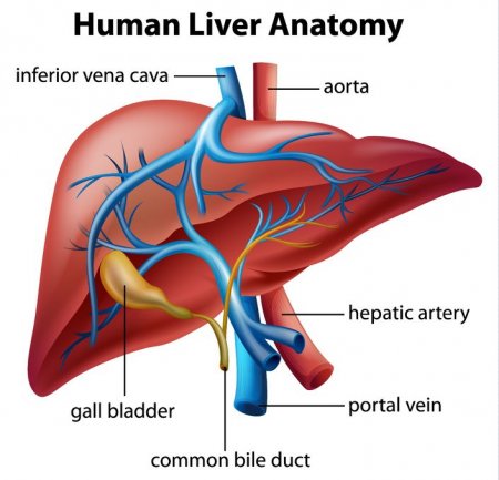
Weighing in at around 3 pounds, the liver is the body’s second largest organ; only the skin is larger and heavier. The liver performs many essential functions related to digestion, metabolism, immunity, and the storage of nutrients within the body. These functions make the liver a vital organ without which the tissues of the body would quickly die from lack of energy and nutrients. Fortunately, the liver has an incredible capacity for regeneration of dead or damaged tissues; it is capable of growing as quickly as a cancerous tumor to restore its normal size and function.
The epiglottis
- Category: Digestive system
- Views: 92451
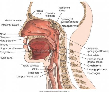
The epiglottis is a flexible flap at the superior end of the larynx in the throat. It acts as a switch between the larynx and the esophagus to permit air to enter the airway to the lungs and food to pass into the gastrointestinal tract. The epiglottis also protects the body from choking on food that would normally obstruct the airway.
The epiglottis is a thin, leaf-shaped structure at the superior border of the larynx, or voice box. In its relaxed position, the epiglottis projects into the pharynx, or throat, and rests just posterior to the tongue. Viewed from the posterior direction, it is shaped like a teardrop with a wide, rounded region at the superior end and a narrow tapered point at its inferior end. The epiglottis is also concave with the lateral edges pointing posteriorly. Two tiny ligaments - the thyroepiglottic and hyoepiglottic ligaments - hold the epiglottis in its resting position in the throat. The thin thyroepiglottic ligament connects the inferior point of the epiglottis to the thyroid cartilage of the larynx, while the hyoepiglottic ligament connects the anterior surface of the superior region to the hyoid bone.
The respiratory system of the head and neck
- Category: Respiratory system
- Views: 94264
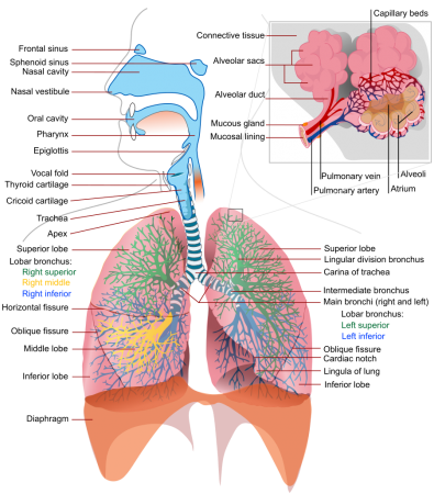
The respiratory system of the head and neck marks the starting point for where oxygen enters the body. The system begins at the nose and mouth where oxygen is inhaled. The areas of the respiratory in the head and neck allow air to flow in and out of the lungs.
The important parts of the respiratory system in the head and neck include the nasal cavity, which processes the airflow on its way through to the lungs. Connected to the nasal cavity is the pharynx that is actually a part of the respiratory and digestive systems. It allows for the passage of both food and air. It lies behind and to the sides of the larynx, or voice box, which forms part of a tube in the throat that carries air to and from the lungs and houses the epiglottis. At rest, the epiglottis is upright and allows air to pass through the larynx and into the rest of the respiratory system. During swallowing, it folds back to cover the entrance to the larynx, preventing food and drink from entering the windpipe. The trachea, or windpipe, allows the head and neck to twist and bend during the process of breathing.
All of these parts in the head and neck play a significant role in directing oxygen to the lungs so that the body can breathe in oxygen.
Physiology of the respiratory system
- Category: Respiratory system
- Views: 12362
Pulmonary Ventilation
Pulmonary ventilation is the process of moving air into and out of the lungs to facilitate gas exchange. The respiratory system uses both a negative pressure system and the contraction of muscles to achieve pulmonary ventilation. The negative pressure system of the respiratory system involves the establishment of a negative pressure gradient between the alveoli and the external atmosphere. The pleural membrane seals the lungs and maintains the lungs at a pressure slightly below that of the atmosphere when the lungs are at rest. This results in air following the pressure gradient and passively filling the lungs at rest. As the lungs fill with air, the pressure within the lungs rises until it matches the atmospheric pressure. At this point, more air can be inhaled by the contraction of the diaphragm and the external intercostal muscles, increasing the volume of the thorax and reducing the pressure of the lungs below that of the atmosphere again.
To exhale air, the diaphragm and external intercostal muscles relax while the internal intercostal muscles contract to reduce the volume of the thorax and increase the pressure within the thoracic cavity. The pressure gradient is now reversed, resulting in the exhalation of air until the pressures inside the lungs and outside of the body are equal. At this point, the elastic nature of the lungs causes them to recoil back to their resting volume, restoring the negative pressure gradient present during inhalation.
The teeth
- Category: Digestive system
- Views: 8150
By the shape the teeth are divided into:
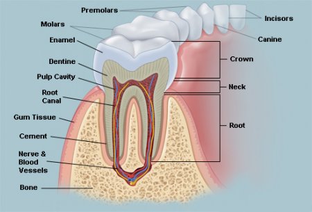
- incisors - have one root, wedge-shaped crowns, particularly upper incisors crown shape has the spatula, lower - bits;
- canines - have one root, crown conical shape;
- premolars: upper premolars have bifurcated root, on the horizontal cut their crown has oval shape; hills are almost identical; lower premolars have one root, on the horizontal cut their crown has round shape, vestibular hill is large, oral - smaller;
- molars: upper molars have three roots (two vestibular and one oral), diamond-shaped crown; lower molars have two roots, square shape of crown.
Adult human has 28-32 permanent teeth. Child has 20 milk teeth.
The teeth are located in the mouth.
The salivary glands
- Category: Digestive system
- Views: 8169
Salivary glands produce saliva.
Salivary glands are divided into small and large.
Small salivary glands located in the mucous membrane of the mouth:
- labial;
- buccal;
- palatine;
- lingual;
- molar.
Large salivary glands:
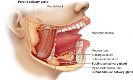
1) parotid gland - paired, is located near the ear, in the mandibular fold. Duct of parotid gland located on chewing muscle, passes through the buccal muscle and opens in the cheek mucosa opposite of 2nd upper molar. Parotid salivary gland - parenchymal organ, anatomical unit of which is the a particle;
2) submandibular gland - paired, located in submandibular triangle. Duct of submandibular gland opens in the proper oral cavity in the sublingual meet. Submandibular gland - parenchymal organ, anatomical unit of which is the a particle;
3) sublingual gland - paired, located in the sublingual crease. Large duct of sublingual gland opens in the sublingual meet, small duct opens directly in sublingual crease. Sublingual gland - parenchymal organ, anatomical unit of which is a particle.
The tongue
- Category: Digestive system
- Views: 23210
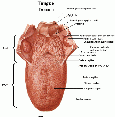
The tongue has a conical shape. The tongue - unpaired body. The tongue is located in the oral cavity.
The outer structure of the tongue
Tongue is divided:
- root of the tongue (radix linguae);
- body of the tongue (corpus linguae);
- apex (tip) of the tongue (apex linguae);
- the back of the tongue (dorsum linguae);
- the lower surface of the tongue (facies inferior linguae).
On the back of the tongue is a blind hole (foramen cecum linguae). From the blind hole aside and ahead goes steam frontier furrow. This furrow divides the tongue into pre-furrow part and after-furrow part.
The internal structure of the tongue
The tongue consists of muscles covered with mucous membrane. The muscles of the tongue are divided into skeletal and own. Skeletal muscle:
- Awl-lingual muscle - starts from subulate process, shifts the tongue up and back;
- Sublingual-lingual muscle - starts from the hyoid bone, the tongue shifts back and down; aside;
- Chin-lingual muscle - starts from chin spine, shifts the tongue ahead.
Development of the oral cavity
- Category: Digestive system
- Views: 7140
In the main part of anterior part of primary intestine develops gill apparatus, which consists of 5 pairs of gill pockets and gill arches. Gill pockets - a protrusion of lateral walls endoderm of the primary intestine. Gill arches - a section of mesenchyme placed between adjacent gill pockets. Primary oral cavity (mouth bay) has the form of cracks, bounded with 5 processes, derivatives of the first pair of gill arches. The upper edge of the mouth bay bounded by unpaired frontal process and by two maxillary processes, the lower edge - by two mandibular processes. Mandibular grow, processes and form the lower jaw, soft parts of the face, chin, lower lip. If mandibular processes do not grow, arises an anomaly - midpoint separation of the lower jaw, lips. Maxillary processes form the upper jaw, palate, lateral parts of the upper lip, cheeks.
Oral cavity
- Category: Digestive system
- Views: 103882
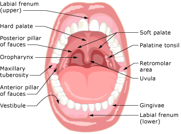
Mouth has 2 parts:
1) oral vestibule
2) proper oral cavity.
1. Vestibule of oral cavity is the space bounded by the upper and lower lips, cheeks (front) and also by teeth and gums (rear).The upper lip is bounded by basis of nose and pair nasolabial sulcus; lower lip is bounded by chin-lip sulcus. Lips are formed by circular muscle of the mouth, which is covered with skin on the outside and by mucous membrane on inside. There are skin part of the lip, intermediate - red border, mucous. In the transition from the upper lip to the alveolar bone of the upper jaw mucous membrane forms a fold - the bridle of upper lip; in the transition from the alveolar part of lower lip to the lower jaw mucous membrane forms a fold - bridle of lower lip. Between the lips is oral cleft. Cheek is bounded in above by zygomatic arch, in bottom – by edge of the lower jaw, in front – by nasolabial sulcus, in behind - by chewing muscles. The basis of cheek is buccal muscle lined with mucous membrane inside, and the outside is covered with skin. In cheek is pronounced subcutaneous fat, which forms the fat body of cheeks.
2. Proper oral cavity is bounded by the upper and lower walls. The upper wall is a palate. There are:
- hard palate, formed by palate bone and mucous membrane that covers the bone palate;
- soft palate, formed by muscles and mucous membrane that covers the muscles.
Splanchnology (SPLANCHNOLOGIA) - the science of internals
- Category: Anatomy
- Views: 84739
The internals - the internal organs that lie in body cavities (mouth, cavity of neck, chest, abdominal, pelvic) and provide the metabolic processes in the organism with the external environment. The internals are combined into organs systems (digestive, respiratory, urogenital).
Development of internals
After 3th week of fetal development, as a result of end of gastrulation are formed three germ layers (ectoderm, mesoderm, endoderm) and axial complex of germs. Mesoderm is divided into:
- dorsal
- intermediate
- ventral.
